
 Fresh Brainz reader Alvin alerted me to a recent review article from Nature Reviews Neuroscience (Lamb et. al 2007) about the evolution of the vertebrate eye, to address the criticism of evolutionary biology by another reader in his blog.
Actually, I thought that the mechanism of eye evolution has already been beaten to death by many researchers, so I'm a tad surprised that there is still any controversy about it today.
But since this review is quite clear and well-organized, I feel that I ought to share it with everyone as a focal point for discussion.
1. Historical background
The review article starts with an exerpt of this quote by Charles Darwin more than 120 years ago:
To suppose that the eye, with all its inimitable contrivances for adjusting the focus to different distances, for admitting different amounts of light, and for the correction of spherical and chromatic aberration, could have been formed by natural selection, seems, I freely confess, absurd in the highest possible degree. Yet reason tells me, that if numerous gradations from a perfect and complex eye to one very imperfect and simple, each grade being useful to its possessor, can be shown to exist; if further, the eye does vary ever so slightly, and the variations be inherited, which is certainly the case; and if any variation or modification in the organ be ever useful to an animal under changing conditions of life, then the difficulty of believing that a perfect and complex eye could be formed by natural selection, though insuperable by our imagination, can hardly be considered real. How a nerve comes to be sensitive to light, hardly concerns us more than how life itself first originated; but I may remark that several facts make me suspect that any sensitive nerve may be rendered sensitive to light, and likewise to those coarser vibrations of the air which produce sound.
The first sentence is often quote-mined by creationists attempting to show that Darwin could not explain eye evolution by natural selection, which isn't what Darwin meant at all.
Still, the eye is undeniably an intricate organ, and to be convinced that it can be produced by stepwise evolution, we need to look deep into the details.
The authors start by providing an overview of the origin of vertebrates, based mainly on the fossil record.

More than 600 million years ago, early organisms already had photoreceptor cells that could be used for shadow detection, in order to escape from predators, and for controlling their circadian rhythm.
They did not have an image forming eye.
Then, our last common ancestor with these primitive bilaterally symmetrical animals (bilateria) diverged from them over 580 million years ago. Animals with a skull (craniates) appeared about 530 million years ago, while animals with vertebrates (vertebrates, duh!) came onto the scene about 500 million years ago.
Sometime between 540 and 500 million years ago, image forming eyes and visual systems came into existence. This corresponds to a period of time called the Cambrian explosion, marked by the rapid evolution of animal body shapes.
Let me point out a few interesting features of the phylogenetic tree shown above.
The authors have highlighted five modern groups of animals that are relevant to the study of the origin of the vertebrate eye.
Obviously they need to include some vertebrate species, and the most diverged species of vertebrate is the lamprey, a jawless fish that has a simple image forming eye.
They also need the nearest outgroup species for comparison. The hagfish is a jawless craniate that doesn't have an image forming eye so it is an appropriate animal to choose.
However, the authors acknowledge that there is molecular evidence that the hagfish could have diverged later from the vertebrates (shown as dotted lines). Until the ancestry of the hagfish is confirmed by genome sequencing, it's a good idea to pick some other animals as outgroup species, such as lancelets and sea squirts.
They have also defined vertebrate eye evolution into 6 stages, shown on the bottom of the chart next to the timeline.
And did you spot the mermaid?
2. Examining the evidence
Next, the authors discussed the structure and function of the eye in those relevant species of animals.
Let me just focus on the hagfish and the lamprey.
The eyes of the hagfish are small and conical. They do not have a lens, an iris, a cornea or any muscles inside or outside the eyes, and they are buried beneath a layer of translucent skin. The retina only have two nuclear layers with no bipolar or amacrine cells, and the photoreceptors directly connect to output neurons. The hagfish doesn't seem to use its eyes for vision, since it's behaviourally almost blind, so the eyes are more likely used as a circadian organ.
In contrast, the lamprey has a camera-like eye, containing a lens and an iris. The retina has three nuclear layers consisting of photoreceptor cell bodies, bipolar cells, horizontal cells, amacrine cells and ganglion cells. Unlike jawed vertebrates however, it has five muscles outside the eye (compared to six) and has no muscles inside the eye (used for focusing in other vertebrates).
Curiously, lamprey larva have eyes that are similar to that of the hagfish - small, buried beneath the skin, and containing a relatively undifferentiated retina. This is a clue that the authors will discuss later.
The authors conclude that the camera-like vertebrate eye already existed in the last common ancestor between jawless and jawed vertebrates, about 500 million years ago.
Is there evidence of how the eye was formed?
Let's zoom into the microscopic details of photoreceptor cells...
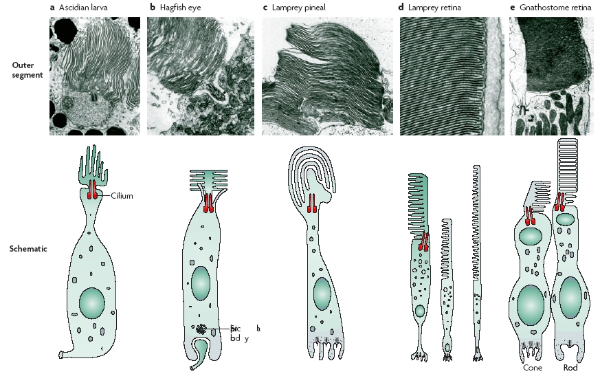
In this figure, five groups of photoreceptors are shown, from sea squirts (left) to mammals (right). You can see a stepwise transition in the alignment of the photopigment membranes, at the top of each cell.
In the sea squirt they are aligned with the axis of the cell. In the hagfish the membranes spread out more laterally. In the lamprey and other vertebrates, the membranes are neatly stacked and aligned perpendicular to the cell axis.
Now let's zoom in even further, all the way down to the molecular level...

There are two types of photoreceptor cells in animals: rhabdomeric (more commonly found in invertebrates) or ciliary (more common in vertebrates).
This figure compares the various light-sensitive receptor proteins, called opsins, found in these cell types. The bottom six opsins are found in the retina of vertebrate eyes.
What is particularly interesting is the middle column labelled "Bovine residue", which refers to key amino acid positions in the opsin genes, using cow rhodopsin as a numbering guide. Similar amino acids are shaded in blue and green.
Notice how there is a transitory, stepwise change at the amino acid level - from opsins that are not used by vertebrates at the top of the chart, down to essential vertebrate retinal opsins at the bottom. This shows that the molecular components of vertebrate photoreceptors were modified from those found in invertebrate photoreceptors.
3. Sequence of events
The authors then discussed the development of the vertebrate eye cup.
They mentioned Karl Ernst von Baer's idea that the developmental stages that an embryo passes through might reflect the evolutionary history of the organism. They know that this idea is overly simplistic (and for some developmental stages, not applicable) but based on a number of observations (for example the lamprey larva mentioned earlier) , they think that it is a good starting point to help formulate testable hypotheses regarding the formation of the retina.

In this figure, the sequence of events in the development of the vertebrate eye is shown. The authors feel that a sequence broadly similar to this might have occurred during the evolution of the vertebrate eye.
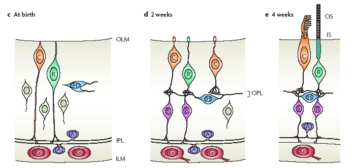
One intriguing clue that support their view at the cellular level is the sequence of events during the development of the retinal circuitry in vertebrates. At birth, photoreceptor cells (C = cone, R = rod) contact the ganglion cell layer (G, output neurons) directly, just like in the hagfish eye.
However as time progresses the photoreceptor projections retract, while bipolar cells (B), horizontal cells (H), and amacrine cells (A) migrate into position.
Eventually the photoreceptors contact output neurons indirectly via other cells in a three-layer retina.
Based on current evidence, the authors propose a multistep sequence of events that result in the formation of the vertebrate eye.

As you can see, it is a long list of many specific details.
More importantly, what's great about putting all this down clearly is that you can then generate some testable predictions to check if the sequence is correct...

These are future experiments that will help refine the certainty and precision of our current understanding of eye evolution.
So you can see that the evolution of the eye is well understood at the anatomical (eye structure), physiological (circuitry and function) and molecular (protein and genetics) levels.
Not as "speculative" and "general" as some people claim.
Would you like to know more?
- Original article
TD Lamb, SP Collin and EN Pugh Jr (2007) Evolution of the vertebrate eye: opsins, photoreceptors, retina and eye cup. Nature Rev Neurosci 8: 960-976


2 Comments:
Dude, you should stick the peer-reviewed blogging icon on this post - it certainly qualifies!
http://bpr3.org/
I've put up the icon. Thanks!
Post a Comment